This summer, New York became the fourth state (after Connecticut, Texas and Virginia) to mandate that women be notified if their mammogram reveals that they have dense breasts -- but what does "dense breasts" actually mean, and how does it affect you?
Mammograms can show whether breasts are made up of mostly dense (meaning milk producing or connective tissue) or fatty tissue. Fatty tissue shows up dark on mammograms, while dense tissue shows up white. The problem is that potentially cancerous spots also show up white, making them harder to spot in dense breasts. The purpose of informing women if they have dense breasts is so they and their physicians can discuss whether additional imaging procedures, such as ultrasound, MRI, or 3D breast tomosynthesis (also known as 3D mammography) would be beneficial to help identify potential problems.
Does having dense breasts mean a reduced chance of surviving cancer if cancer is discovered? The answer, according to a recent study, is no.
The study, published in the Journal of the National Cancer Institute, followed over 9,000 breast cancer patients and found that patients with dense breasts were no more likely to die from breast cancer than those with less dense breasts. So, while tumors in dense breast tissue may be harder to detect, once detected they are no more aggressive or harder to treat than tumors in less dense breasts.
What does this mean to you? If you are notified that you have dense breasts, speak to your physician about whether additional testing might be useful for you, such as 3D breast tomosynthesis. Patients who are classified as having dense breasts benefit the most out of this new technology. Ultimately, take comfort in knowing that breast cancer patients with dense breasts do not have worse outcomes than those without dense breasts.
For more information about the study, visit http://jnci.oxfordjournals.org/content/104/16/1218.abstract
Friday, August 24, 2012
Monday, August 20, 2012
Preventing Misdiagnosis in Breast Cancer
How common are lab errors? According to one study, 1 in 100 women (1%) may receive the wrong diagnosis due to a lab mix-up. With about 1.6 million women having breast biopsies each year, simple math shows that an alarming number may be misdiagnosed. How do we make sure that NEVER happens with our patients? Read on...
Question: I will be having a breast biopsy in a week, and I'm very concerned about something. I am worried that my sample might get mixed up with the sample of someone else, and that I might get a false positive that way. How do you make sure that samples don't get mixed up and that I get the proper results?
Answer: At Zwanger-Pesiri Radiology, we understand your concerns. Your fear is one that is shared by many people, and for good reason. Please rest assured that at ZPR we ensure that you get the right results by using the know error® breast biopsy system.
The system is very simple and painless, and uses DNA technology to match positive samples to patients. Here's how it works in a nutshell: Before your biopsy, a technologist will take a swab of the inside of your cheek. This gathers a sample of your DNA. As you probably know, DNA is individual to each person. No two people have the same DNA.
After your biopsy, all the samples, and your swab are carefully labeled and sent for analysis. In the event that your biopsy result comes back positive, the DNA from your sample is matched with the DNA from your cheek swab. You can have peace of mind that there is no way that you will get a result that isn't yours.
This is just one of the ways that ZPR strives to ensure patient safety and high standards for all of our patients across Nassau and Suffolk counties of Long Island. If you have any questions or concerns that you would like addressed, please contact us.
Question: I will be having a breast biopsy in a week, and I'm very concerned about something. I am worried that my sample might get mixed up with the sample of someone else, and that I might get a false positive that way. How do you make sure that samples don't get mixed up and that I get the proper results?Answer: At Zwanger-Pesiri Radiology, we understand your concerns. Your fear is one that is shared by many people, and for good reason. Please rest assured that at ZPR we ensure that you get the right results by using the know error® breast biopsy system.
The system is very simple and painless, and uses DNA technology to match positive samples to patients. Here's how it works in a nutshell: Before your biopsy, a technologist will take a swab of the inside of your cheek. This gathers a sample of your DNA. As you probably know, DNA is individual to each person. No two people have the same DNA.
After your biopsy, all the samples, and your swab are carefully labeled and sent for analysis. In the event that your biopsy result comes back positive, the DNA from your sample is matched with the DNA from your cheek swab. You can have peace of mind that there is no way that you will get a result that isn't yours.
This is just one of the ways that ZPR strives to ensure patient safety and high standards for all of our patients across Nassau and Suffolk counties of Long Island. If you have any questions or concerns that you would like addressed, please contact us.
Wednesday, August 1, 2012
Healthy Eating for Kids
Our recent newsletter covers Kids and Radiology. This week's blog post will talk about how to get and keep kids eating healthy. If you don't get our monthly e-newsletter, and you'd like to, subscribe here.
Children can be notoriously picky eaters and getting them to try new foods (especially vegetables) and eat a balanced diet, can sometimes be challenging. Here are some tips for encouraging healthy eating habits in kids:
Eat the Rainbow. Brightly colored fruits and vegetables, e.g., carrots, sweet potatoes, red peppers, blueberries, strawberries and tomatoes are healthier than "white" vegetables such as potatoes. Encourage kids to eat a rainbow of foods each day.
Go for Organic. Young children are more sensitive to pesticides and chemicals used in plant growth. If organic produce isn't too costly, it's well worth it. To see which fruits and vegetables are best to buy organic and which are safe to buy non-organic, visit the Environmental Working Group's Shopper's Guide.
Play with Food. Letting kids play with their food -- i.e., make their own creations or make decorative foods -- is a great way to incorporate different veggies. Who doesn't like broccoli "trees" or carrot "swords" or "boats" made out of zucchini or cucumber?
Hide the Veggies. For kids who are resistant to eating vegetables, try disguising them. Zucchini bread, carrot cake, and bran muffins are all easy ways to slip some nutrition into what seems to be a treat. Also consider adding shredded carrots or zucchini into meatloaf, and mixing vegetables into pasta.
Healthy Substitutions. Instead of cookies or candy, give kids bowls of berries, cherries, or mixed raisins and nuts for snacks. Instead of french fries, try oven roasted sweet potato wedges. Instead of iceberg lettuce, try a darker green like spinach or mixed baby greens. Use fresh vegetables whenever possible, and frozen rather than canned.
Let them Cook. When children are involved in the cooking, they will be more likely to try the finished product. Let kids take part in food preparation (supervised of course) and they may be willing to try foods they never would have tried before.
Children can be notoriously picky eaters and getting them to try new foods (especially vegetables) and eat a balanced diet, can sometimes be challenging. Here are some tips for encouraging healthy eating habits in kids:
Eat the Rainbow. Brightly colored fruits and vegetables, e.g., carrots, sweet potatoes, red peppers, blueberries, strawberries and tomatoes are healthier than "white" vegetables such as potatoes. Encourage kids to eat a rainbow of foods each day.
Go for Organic. Young children are more sensitive to pesticides and chemicals used in plant growth. If organic produce isn't too costly, it's well worth it. To see which fruits and vegetables are best to buy organic and which are safe to buy non-organic, visit the Environmental Working Group's Shopper's Guide.
Play with Food. Letting kids play with their food -- i.e., make their own creations or make decorative foods -- is a great way to incorporate different veggies. Who doesn't like broccoli "trees" or carrot "swords" or "boats" made out of zucchini or cucumber?
Hide the Veggies. For kids who are resistant to eating vegetables, try disguising them. Zucchini bread, carrot cake, and bran muffins are all easy ways to slip some nutrition into what seems to be a treat. Also consider adding shredded carrots or zucchini into meatloaf, and mixing vegetables into pasta.
Healthy Substitutions. Instead of cookies or candy, give kids bowls of berries, cherries, or mixed raisins and nuts for snacks. Instead of french fries, try oven roasted sweet potato wedges. Instead of iceberg lettuce, try a darker green like spinach or mixed baby greens. Use fresh vegetables whenever possible, and frozen rather than canned.
Let them Cook. When children are involved in the cooking, they will be more likely to try the finished product. Let kids take part in food preparation (supervised of course) and they may be willing to try foods they never would have tried before.
Thursday, July 26, 2012
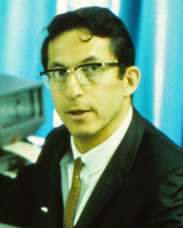
Dr. Robert Ledley -- Inventor of the CT Scan
ZPR mourns the passing of Dr. Robert Ledley, the inventor of the first Computed Tomography (CT) scanner that was capable of imaging any part of the body. He passed away on Tuesday, July 24th, at the age of 86.
As a young man, Ledley wanted to study physics. His practical parents told him he could, providing he also became a licensed dentist. His parents wanted to be sure he could make a good living, so Ledley studied physics but also became a dentist. In the late 1950s, he became interested in computers and began focusing on how computers could assist in solving biomedical problems. In 1960, Ledley founded the National Biomedical Research Foundation, a nonprofit organization dedicated to promoting the use of computing methods among biomedical scientists. By the early 1970s, he began his work on CT scanning.
His first machine was the Automatic Computerized Transverse Axial (ACTA) scanner, which was the first one capable of scanning the entire body. The original prototype of the ACTA scanner is at the Smithsonian Institution, and Ledley was inducted into the National Inventors Hall of Fame in 1990 and awarded the National Medal of Technology and Innovation by President Clinton in 1997.
Prior to Dr. Ledley's invention, radiologists had limited resources -- primarily, traditional X-rays. CT scanning provides much higher resolution images than X-rays, as well as being able to produce cross-sectional, 3D images. The invention of the CT scanner has changed how physicians diagnose and treat diseases.
ZPR acknowledges Dr. Ledley's great contribution to our field and is proud to offer the latest in CT technology, including the 256-slice CT-Flash scanner which exposes patients to 75% to 90% less radiation than most other scanners.
As a young man, Ledley wanted to study physics. His practical parents told him he could, providing he also became a licensed dentist. His parents wanted to be sure he could make a good living, so Ledley studied physics but also became a dentist. In the late 1950s, he became interested in computers and began focusing on how computers could assist in solving biomedical problems. In 1960, Ledley founded the National Biomedical Research Foundation, a nonprofit organization dedicated to promoting the use of computing methods among biomedical scientists. By the early 1970s, he began his work on CT scanning.
His first machine was the Automatic Computerized Transverse Axial (ACTA) scanner, which was the first one capable of scanning the entire body. The original prototype of the ACTA scanner is at the Smithsonian Institution, and Ledley was inducted into the National Inventors Hall of Fame in 1990 and awarded the National Medal of Technology and Innovation by President Clinton in 1997.
Prior to Dr. Ledley's invention, radiologists had limited resources -- primarily, traditional X-rays. CT scanning provides much higher resolution images than X-rays, as well as being able to produce cross-sectional, 3D images. The invention of the CT scanner has changed how physicians diagnose and treat diseases.
ZPR acknowledges Dr. Ledley's great contribution to our field and is proud to offer the latest in CT technology, including the 256-slice CT-Flash scanner which exposes patients to 75% to 90% less radiation than most other scanners.
Wednesday, July 18, 2012
ZPR and You: The Patient Portal
Zwanger-Pesiri Radiology is always looking for new ways to improve service for our patients. One recent, and exceptionally useful, improvement was the launch earlier this year of our Patient Portal. The Patient Portal gives YOU access to your reports and information via your computer. Using the Patient Portal you can:
- View your exam results
- See a record of all your procedures and appointments
- Make payments and view your balance
- View upcoming appointments
The Patient Portal is just one of the ways that ZPR is helping you to have more control of your own health care. Stay tuned for upcoming blog posts that will cover other "ZPR and You" topics.
Frequently Asked Portal Questions
Wednesday, July 11, 2012
Radiology Specialties
In previous blog posts, we've already established that radiologists are highly trained physicians, and we've looked at some of the things that radiologists do. In this week's post, we will look at some radiology specialties.
Radiologists are specialists at diagnosing disease and injury using medical imaging techniques. But there are also many subspecialties of radiology, including:
- Neuroradiology which focuses on the diagnosis of brain, spinal cord, head, neck, and vascular problems. Some diagnostic tests that neuroradiologists perform include: sinus CT for evaluation of inflammatory disease (sinusitis), neck CT to assess the larynx, lymph nodes and salivary glands, CT and MR angiograms to detect aneurysms, and MRI to evaluate brain tumors, multiple sclerosis, strokes, and neck and back pain.
- Body Imaging which involves studies of the chest, abdomen and pelvis, including: prostate MRI, cardiac MRI, abdominal CT to identify kidney and ureter stones, lung CT and many more. Ultrasound may be performed to evaluate abdominal or pelvic pain, vascular disorders and for obstetrics.
- Breast Imaging which uses digital mammography, positron emission mammography, 3D breast tomosynthesis, breast MRI, and breast specific gamma imaging to diagnose and stage breast cancer. Office-based biopsies are also performed, when necessary.
- Musculoskeletal Imaging, which as the name implies, identifies issues with muscles, bones and joints using MRI and CT. Radiologists who specialize in this field also use MR arthrography to help identify the cause of joint pain, most often in the shoulder.
- Nuclear Medicine which diagnoses diseases using a radioactive agent -- called a radiopharmaceutical -- along with imaging technology. Nuclear medicine specialists are physicians who have specialized and become board certified in nuclear medicine. Some radiologists also have additional specialized training in nuclear medicine.
Tuesday, July 3, 2012
What does a radiologist do?
Now that we've established that radiologists are highly trained physicians, it's time to address the question of what radiologists do. Because they operate more "behind the scenes," patients are sometimes confused about the role of a radiologist.
Q: When I come in for my MRI, the radiologist is the one who is performing the test, right?
A: Wrong! As a general rule, the people who conduct the tests are radiologic technologists. Radiologic technologists have been specially trained to operate the equipment, and they operate under the supervision of radiologists, but radiology techs are not doctors.
Q: Then what is the role of the radiologist?
A: Radiologists diagnose disease and injury using medical imaging technologies -- such as MRIs or CT scans. Just as your regular physician might diagnose your health problem by examining your body, radiologists are specially trained to interpret results of imaging studies and then convey this information to your referring physician. Radiologists also commonly work with referring physicians to help choose the best imaging technique for each patient's specific issue.
Q: So the radiologist works with my referring doctor?
A: Yes, your radiologist plays an important role in your health by acting as an expert consultant to your referring physician. Besides helping him or her choose the proper imaging exam, your radiologist will also interpret the resulting medical images and use those results to recommend further scans or treatments if necessary.
Subscribe to:
Posts (Atom)